Fluorescence Microscopy
3.5.16.
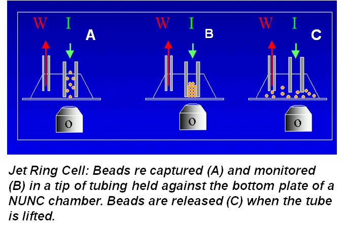
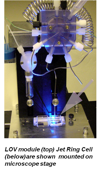
BI fluorescence microscopy, originally designed in conventional SI format (Hodder 1999, Scampavia 1999), was later adopted to the LOV platform, (Schultz 2000) in combination with a Jet Ring Cell. This cell, used to capture the beads, is formed by a stainless steel tubing mounted perpendicularly to a quartz window of a NUNC chamber, and placed on the stage of an inverted microscope. This approach offers numerous advantages as compared to manual methods, or even automated continuous perfusion of cells adherently mounted on a microscope slide since:


- microfluidic control secures reproducibility of the rate of stimulant addition and its perfusion of cellular receptors.
- cellular responses are monitored in reaol time, while the stimulant is introduced, thus providing temporal information on the onset, rate and duration of the response.
- automated renewal of cell population for each experiment eliminates memory effects.
biological variability is eliminated within a series of measurements.
While fluorescence microscopy played an important role in development of BIS for cellular assays, it has been recently replaced by fluorescence measurements using optical fibers that define the flow cell in the LOV module using a photomultiplier as detector and high intensity LED as well as appropriate filters. This section is therefore included to assist the reader in perusing the following slides.